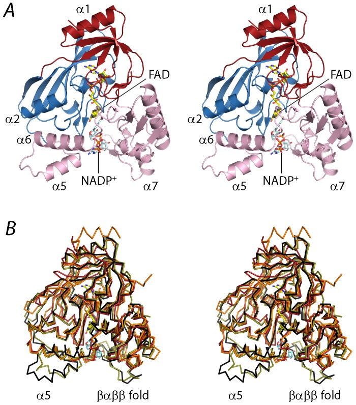
Figure 3. Overall structure of the ternary complex of PaMurB with FAD and NADP+.
A. Stereo view of the crystal structure of PaMurB with bound FAD and NADP+. The enzyme is shown in cartoon representation and comprises FAD-binding domain I (red) and domain II (blue), and the substrate-binding domain III (pink). FAD and NADP+ are shown as yellow and cyan stick models, respectively. NADP+ occupies the channel between the two lobes of domain III (in this view: left, lobe 1; right, lobe 2). Relevant secondary structure elements are labeled. B. Stereo view of the superimposition of the Cα traces of EcMurB (olive green), SaMurB (orange) and TcMurB (red) against PaMurB (black). PaMurB and EcMurB display highly similar structures in their respective NADP- and UNAGEP-bound complexes. In domain III, type II MurB enzymes lack the tyrosine loop preceding helices α4 and α5, as well as the protruding βαββ fold on lobe 2.

