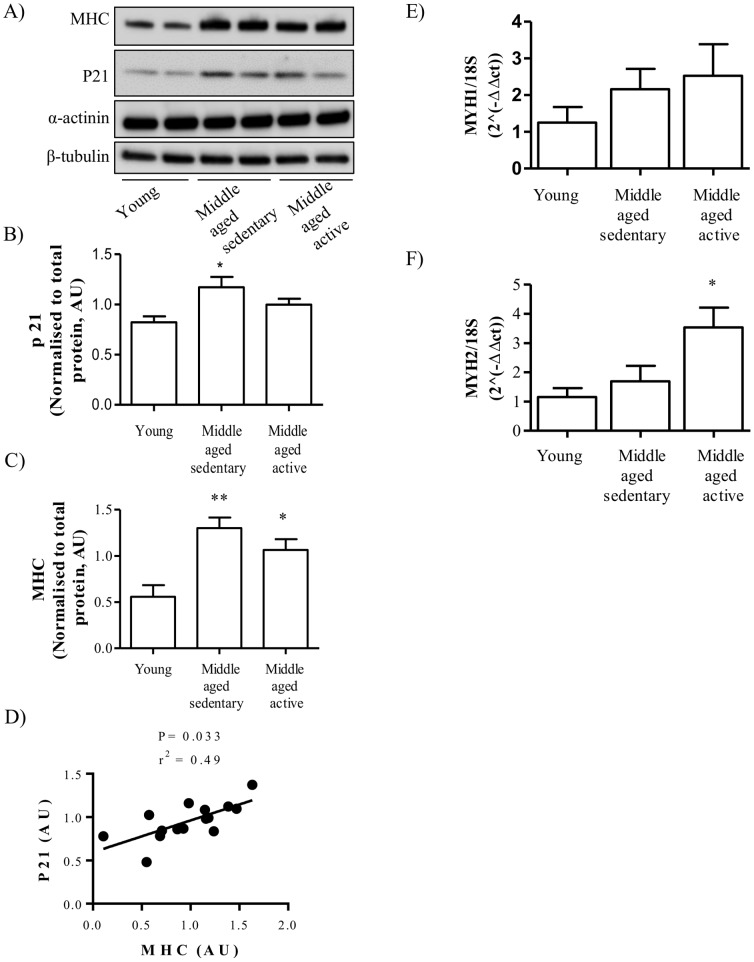
Figure 1. Effect of aging and physical activity on cell cycle and myogenic differentiation.
Satellite cells were isolated from vastus lateralis biopsies from young or middle-aged volunteers: sedentary or active. Cells were grown in culture until mature myotubes were formed. (A) Lysates were immunoblotted to assess the total protein amount of MHC, p21 (Waf1/Cip1) and α-actinin. Equal gel loading was ascertained by immunoblotting with an antibody against β-tubulin. Protein expression of (B) p21 and (C) MHC were quantified and expressed as arbitrary units. (D) Protein quantification of MHC was correlated to protein expression of p21 for all groups. Expression of (E) MYH1 and (F) MYH2 were measured by qPCR and using the delta CT method (AU). Values shown are the mean ±S.E.M from cells from 5 individuals for each group. An asterisk denotes a significant difference from young (*P<0.05, **P<0.01).

