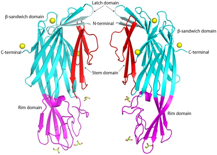
Figure 1. Two views at 180°C of the C. perfringens Delta toxin structure in cartoon representation.
Latch domain, β-sandwich domain, Stem domain and rim domain are colored in pale cyan, cyan, red and magenta, respectively. Glycerol molecules are shown as sticks. Zinc molecules are depicted as yellow balls. Figures 1–4 are produced with PyMol.

