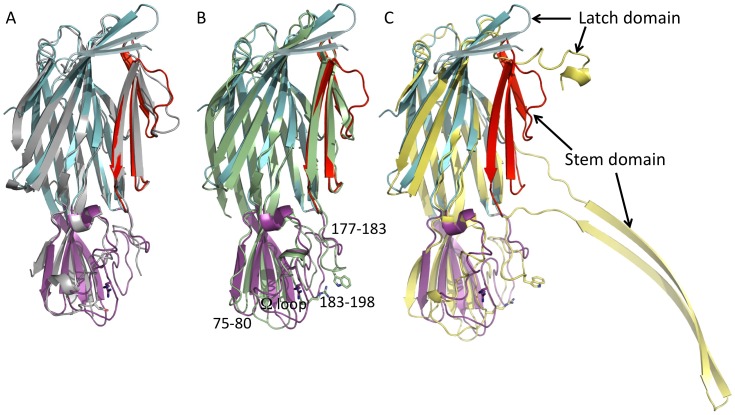
Figure 2. Superposition of C. perfringens Delta toxin structure with γHL-Hlg2 in grey (PDB ID: 3B07) (A), with γHL-LukF in pale green (PDB ID: 3B07) (B) and with a monomer of the αHL of S. aureus in yellow (PDB ID: 7AHL) (C).
Colors for the different domains of C. perfringens Delta toxin have been kept as for Fig. 1. The conserved Arginine and Tryptophan associated with phospholipid binding are shown as sticks. Loops in the rim domain that differ in the various toxins are identified in (B) by their residue numbers in Delta.

