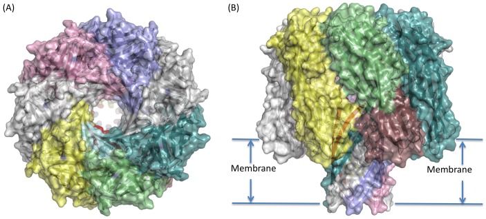
Figure 3. Model of the Delta toxin heptameric pore shown in cartoon representation with a semi-transparent surface.
Chain A is coloured cyan for the latch domain, pale green for the β-sandwich, red for the stem and raspberry for the stem domains. Remaining chains shown in single colour (pale teal, grey, lilac, pink white and yellow). (A) Top (looking down at extra-cellular face) and (B) side-view, indicating possible membrane location.

