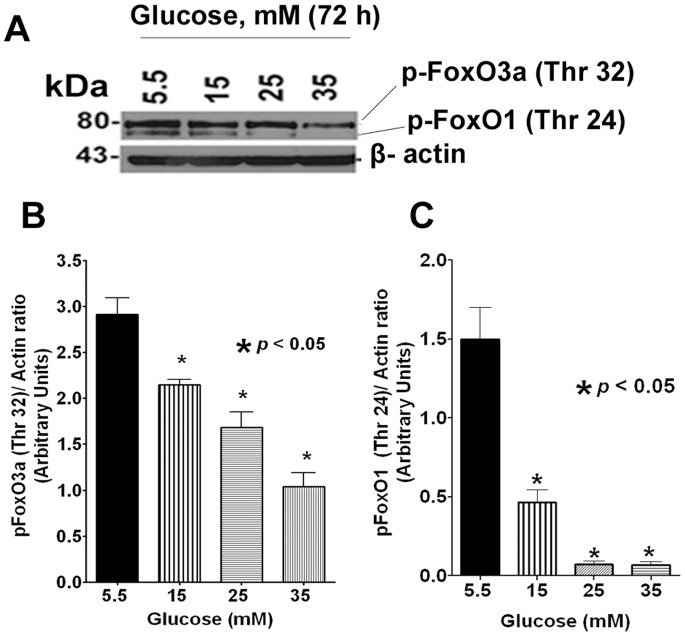
Figure 3. Western blot analysis of phospho-FoxO1 (Thr 24)/phospho-FoxO3a (Thr 32).
A. A typical Western blot image of pFoxO1 (Thr 24)/pFoxO3a (Thr 32) protein expression after glucose treatment for 72 h. B and C. Densitometric analysis of the Western blot from three independent experiments. Data showed that both pFoxO1 (Thr 24) and pFoxO3a (Thr 32) were significantly dephosphorylated under hyperglycemic conditions in a dose-dependent manner. The β-actin was shown as loading control.

