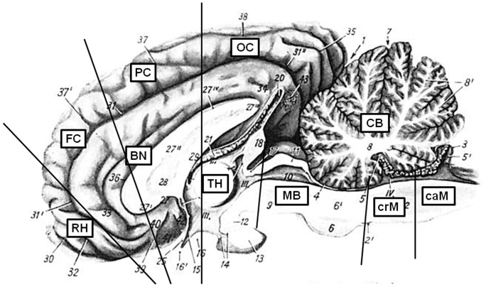
Figure 1. Analysed brain regions.
This picture was modified after Nickel/Schummer/Seiferle [31]. Ten brain regions were analysed: CB – cerebellum, MB – midbrain including pons, crM – cranial medulla oblongata, caM – caudal medulla oblongata, RH – rhinencephalon (olfactory bulb), FC – frontal cortex, PC – parietal cortex, OC – occipital cortex, BN – basal nuclei, TH – thalamus. The medulla oblongata was separated into three regions, but due to the limited amount, only the cranial and caudal medulla could be investigated in detail.

