
Figure 8. The 3D meibomian gland of subject C (67 years old, male) with severe MGD (meiboscore grade 3).
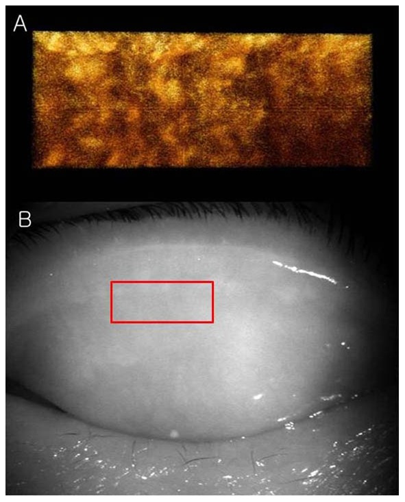
A: The 3D images showed a few meibomian glands with a normal grape-like pattern. We could not find definite acini attached to the central ducts. B: These findings are consistent with the noncontact infrared meibography, which showed nearly complete loss of the meibomian glands.