
Figure 10. The 3D meibomian gland of subject E (a 31-year-old male) with severe MGD (meiboscore grade 3) and graft-versus-host disease.
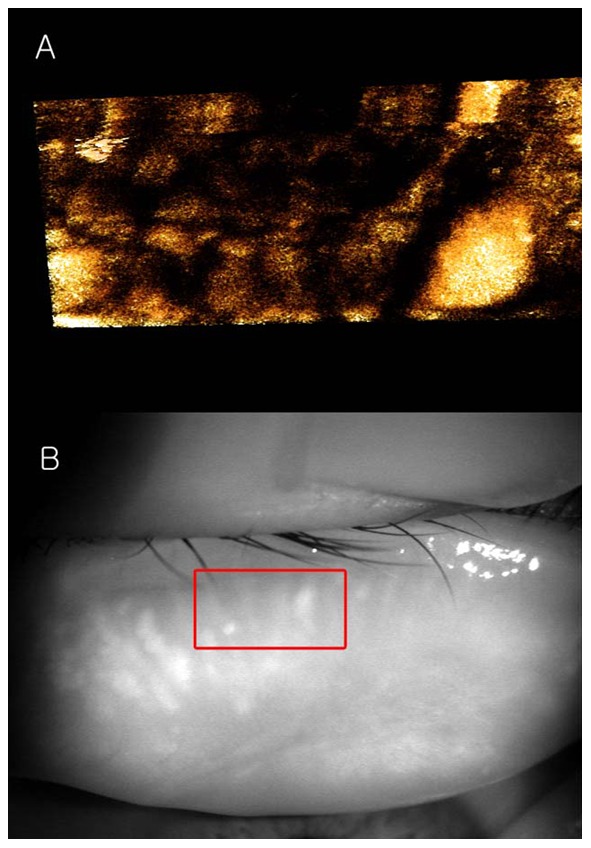
A: We found few meibomian glands with normal morphology, but observed spindle-shaped or globular structures instead; B: The 3D meibomian gland image was consistent with the meibomian glands in the rectangle in the infrared image.