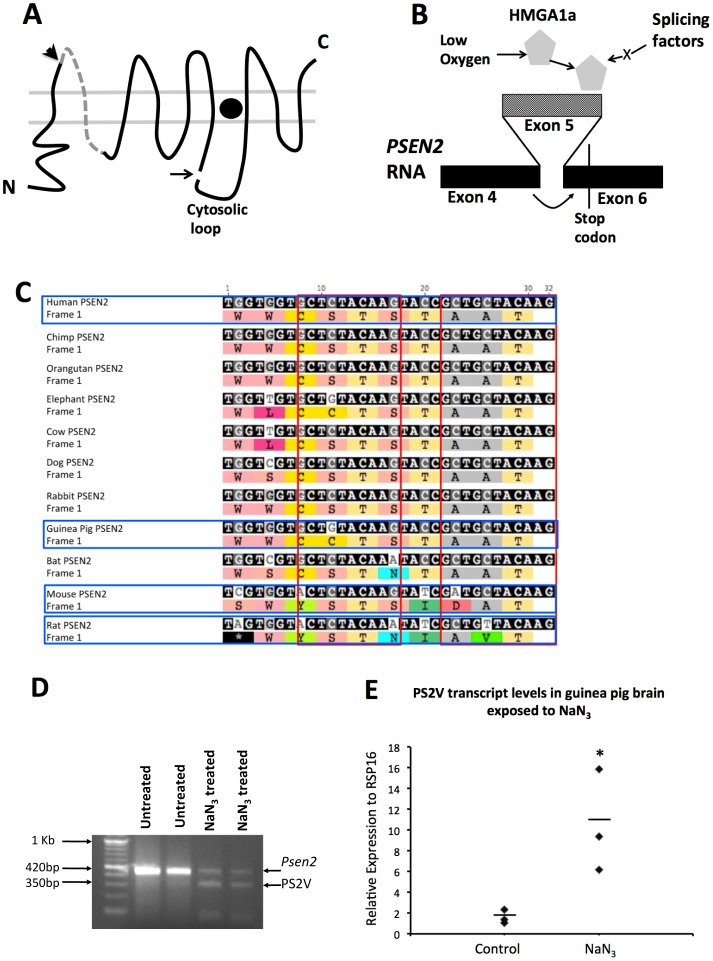
Figure 3. Formation of the PS2V Transcript.
A) Presenilin structure in lipid bilayers: Arrowhead indicates boundary between protein sequences derived from exon 4 and 5. Dashed line indicates sequence from exon 5. Arrow indicates endoproteolysis site. Filled circle indicates γ-secretase catalytic site. B) PS2V forms when HMGA1a is expressed and binds to exon 5 (lighter shading) of PSEN2 RNA causing ligation of exon 4 to exon 6 and ORF termination. C) Nucleotide sequence alignment of the 3′ end of exon 5 in human PSEN2 RNA (with corresponding encoded residues) and the cognate exon of other species. Red boxes enclose sequences aligned with the HMGA1a-binding sites in human PSEN2 RNA. D) mRNA from guinea brains exposed to control media or to media containing NaN3 followed by RT-PCR analysis using primers amplifying cDNA spanning exons 3 to 7 of Psen2. In untreated samples a prominent ∼420 bp band is observed. In NaN3 treated samples an additional ∼350 bp band is evident representing the cDNA fragment predicted from exclusion of the exon 5 sequence (PS2V). E) qPCR using a primer spanning the exon 4/6 junction PS2V cDNA showed up-regulation of PS2V mRNA in samples treated with NaN3.

