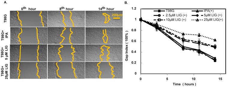
Figure 2. The addition of LIG to T98G cell cultures reduced migration capacity.
A) Images of the wound-like gap closure process of T98G cells under mock, 0.5% isopropanol (IPA), 5 µM LIG, and 25 µM LIG (top to bottom) are presented at the 0th, 8th and 14th hour. The yellow lines indicate the approximate boundary between cell-inhabited and cell-free (central) regions of each image. Scale bar: 200 µm. B) The result of a quantitative analysis of the gap closure of T98G cells under no treatment, 0.5% IPA, and 4 concentrations (2.5, 5, 15 and 25 µM) of LIG treatment is shown. The gap index is the percentage of original wound gap that remains cell-free. The data are the mean gap index results of 6 experiments with error bars representing the standard deviation (also refer to Fig. S1).

