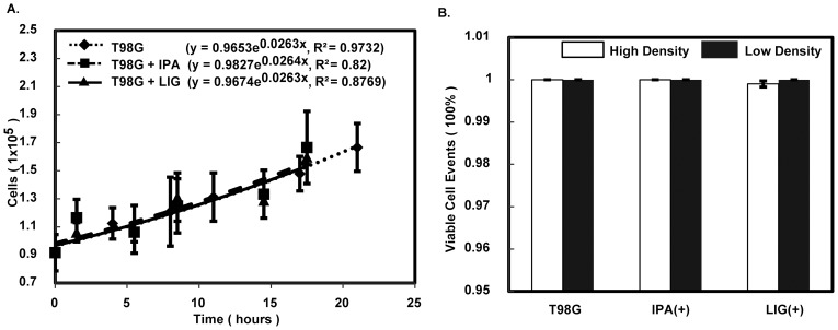
Figure 3. Neither cell proliferation nor apoptosis explain the attenuation of wound-like gap closure in the presence of LIG.
A) Comparative growth of T98G cells under no treatment, 0.5% IPA and 5-µM LIG treatment is shown. Each population of T98G cells was counted every 3 hours for 24 hours. The average number from 9 assessments at each time point was fit to an exponential curve to obtain the growth rate under each treatment condition. Error bars represent the standard deviation. B) The result of apoptosis assays for both high and low density of T98G cells is shown. The bars represent the average between two measurements of viable cell percentages under no treatment, 0.5% IPA, and 5-µM LIG treatment, respectively.

