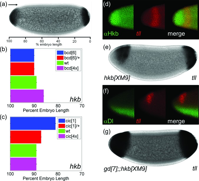
Figure 3.
Anterior repression of tll depends on Hkb and Dl. (a) Quantification of the boundary of anterior hkb. (b) and (c) Quantified boundaries of the anterior hkb in embryos with different levels of Bcd (b) and Cic (c). (d) Averaged boundaries are shown with error bars indicating s.e.m. The numbers of embryos used in the analysis are N = 101 (bcd[6]), N = 64 (bcd[6]/+), N = 52 (wt), N = 67 (bcd[4x]), N = 39 (cic[1]), N = 103 (cic[1]/ +), and N = 41 (cic[4x]). (e) Dorsal view of an embryo stained with Hkb protein (green) and tll transcript (red). The pattern of Hkb closely matches with the repression domain of tll. (e) Pattern of tll in Hkb mutant embryo. (f) Lateral view of an embryo stained for Dl protein and tll mRNA. (g) tll mRNA pattern in an embryo that lacks Dl signaling and Hkb protein.

