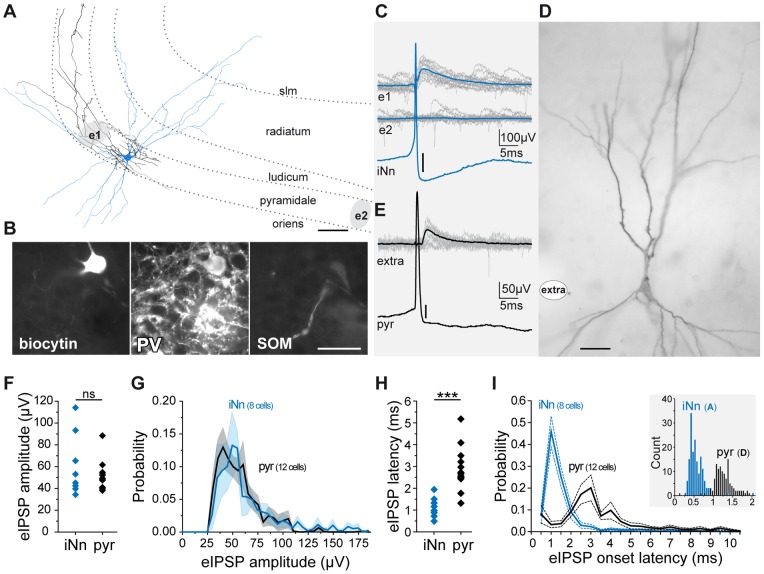
Figure 2. eIPSPs induced by single interneurons and pyramidal cells in CA3.
A–E. eIPSPs triggered by the discharge of single action potentials by a CA3 parvalbumin positive basket cell and by a pyramidal cell. Both neurons were recorded intracellularly in the CA3 pyramidal layer of rat slices and filled with biocytin. A. Camera lucida drawing of the axon (black) and dendrites (blue) of the interneuron and position of the extracellular electrodes (grey areas e1 and e2); scale bar 25 µm; dotted lines indicate the borders of the hippocampal strata: lacumosum-moleculare (slm), radiatum, lucidum, pyramidale and oriens. B. Immunohistochemical characterization of the same cell; scale bar 50 µm. Note perisomatic projection (A), parvalbumin positive (B, PV) and somatostatin negative (B, SOM) labelling. C. A single action potential from this cell (c bottom, averaged trace, scale bar 3 mV) was associated with an eIPSP in the CA3 pyramidal layer at the recording site corresponding to the interneuron projection area (e1, upper traces, 15 superimposed sweeps) but not at another recording site outside this area (e2, lower traces). D. photograph of the recorded and biocytin labeled pyramidal cell; scale bar 20 µm; white spot, extracellular recording site. Note the thorny excrescences on the proximal apical dendrites. E. eIPSPs (upper traces, 15 superimposed sweeps) triggered by the discharge of single action potentials (bottom, averaged trace, scale bar 10 mV) by the morphologically identified CA3 pyramidal cell shown in D. F–I. Amplitudes and latencies of eIPSPs evoked by the discharge of single spikes from individual interneurons (iNn) and pyramidal cells (pyr) recorded intracellularly. F. Average amplitude (one point per presynaptic neuron) of the evoked eIPSPs. G. Distribution of the amplitudes (mean±SEM) of the evoked eIPSPs. H. Average latencies (one point per presynaptic neuron) of the evoked eIPSPs. I. Distribution of the latencies (mean±SEM) of the evoked eIPSPs. Note that the distribution of interneuronal spikes to eIPSP latencies suggests monosynaptic transmission, while the longer latencies of eIPSPs evoked by pyramidal cells spikes suggest disynaptic transmission. Inset shows example histograms of eIPSP latencies for the interneuron shown in A (iNnd, blue bars) and for the pyramidal cell shown in D (pyr, black bars).

