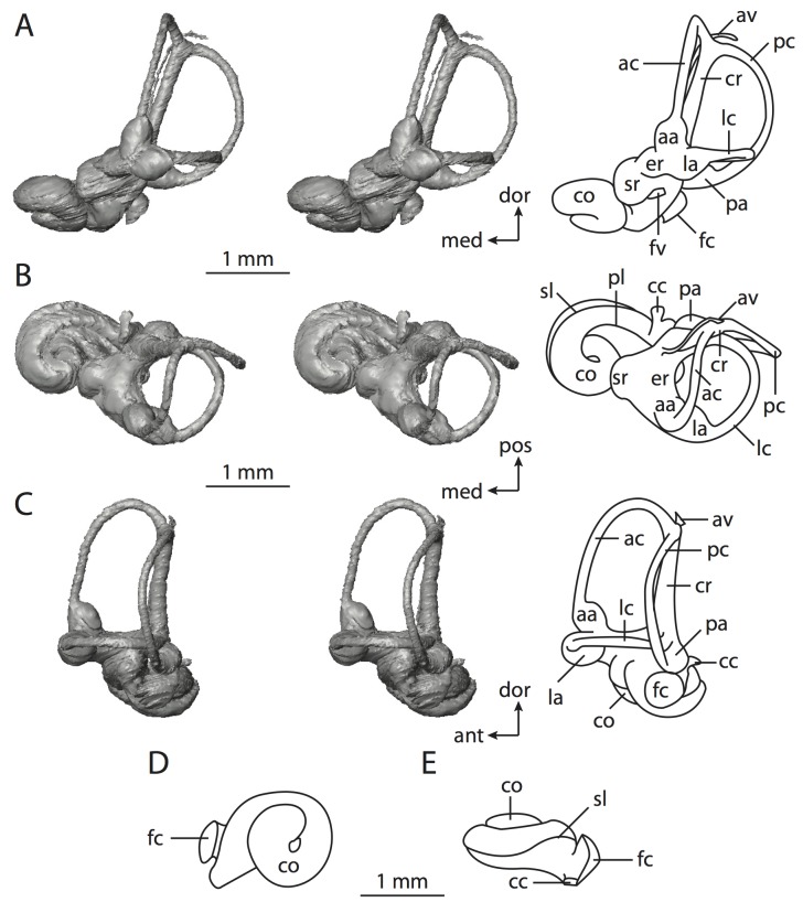
Figure 52. Bony labyrinth ofSorex monticolus.
A , stereopair and labeled line drawing of digital endocast in anterior view; B, stereopair and labeled line drawing of digital endocast in dorsal view; C, stereopair and labeled line drawing of digital endocast in lateral view; D, line drawing of cochlea viewed down axis of rotation to display degree of coiling; E, line drawing of cochlea in profile. Abbreviations listed at the end of the Materials and Methods section.

