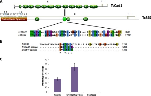
FIGURE 4.

TcSSS contains cadherin repeats and a putative binding epitope homologous to other Bt cadherin functional receptors, which enhances Cry3Ba toxicity in Tc larvae. A, schematic representation of TcCad1 and TcSSS receptors secondary structure obtained with Motif Scan showing the Clustal alignment corresponding to an identified pattern in TcSSS and TcCad1 sequences using PRATT 2.1 (33). The extracellular (E), transmembrane (T), and intracellular (I) domains and numbered cadherin repeat regions (CR1–CR12) are illustrated. B, clustal alignment of the previously described cadherin Bt toxin binding epitopes in M. sexta MsBtR1 (1416GVLTLNIQ1423) and in T. molitor TmCad1 (1359GDITINFE1366) (7, 36) and residues 1103–1150 of the TcSSS sequence corresponding to the homology region in TcCad1 identified using PRATT. In this TcSSS fragment, a putative binding epitope (1115GSATVELK1122) was found. C, enhancement of Cry3Ba toxicity to Tc larvae by a 29-mer peptide (PepTcSSS) spanning amino acids 1110–1138, containing the identified putative binding epitope in TcSSS protein.
