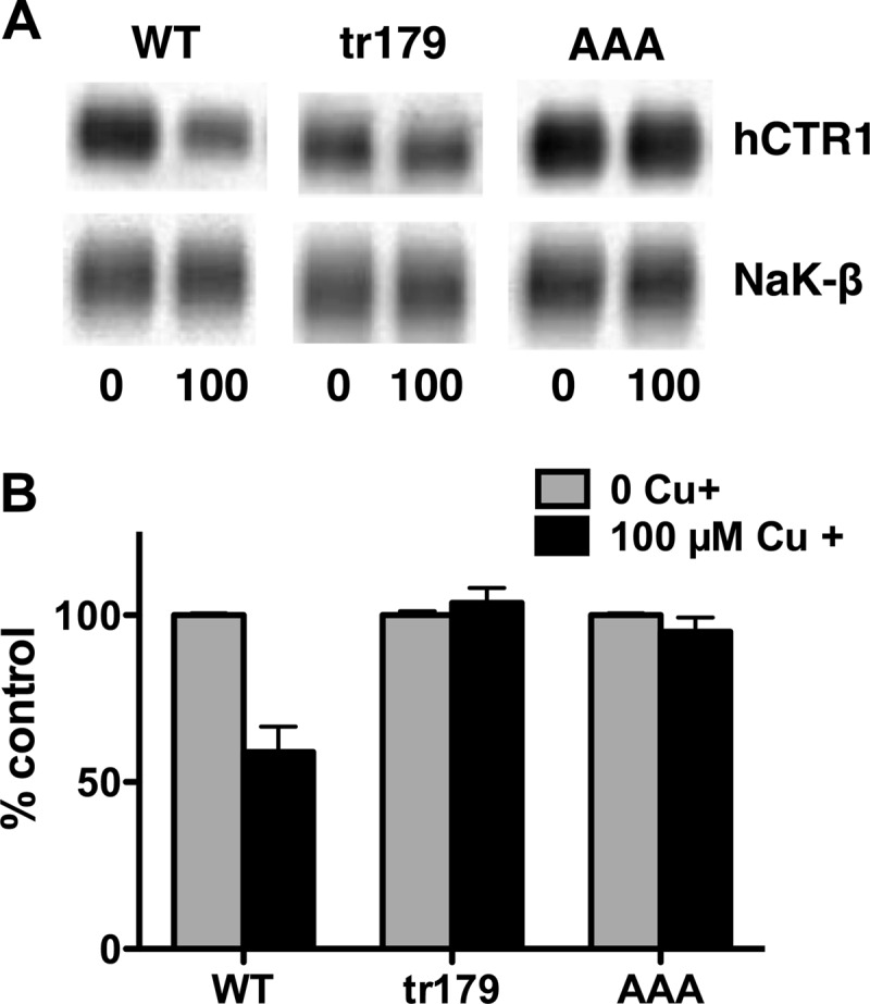
FIGURE 9.

Effect of extracellular copper on cell surface hCTR1 in WT and C-terminal mutant expressing cells. Cells were treated with 100 μm copper for 1 h prior to being biotinylated. A, Western blots of biotinylated proteins were probed with anti-FLAG (overexpressed hCTR1 proteins) and anti-Na,K-ATPase β1-subunit antibodies. B, quantification of Western blots. Relative amount hCTR1 in cells treated in high copper (black bars) is given as the percentage of the level in cells with no added copper in the media. Values are the mean of three separate experiments ± S.D.
