FIGURE 7.
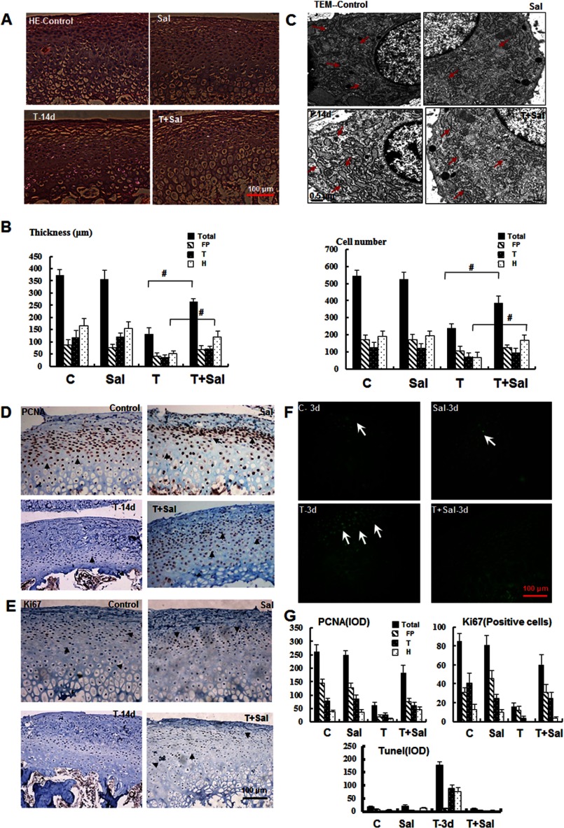
Inhibition of ERS alleviates mandibular cartilage thinning. A, H&E staining of the mandibular cartilage from samples treated as indicated for 14 days (magnification ×100). Sal, salubrinal. B, quantification of the results shown in A for the cartilage thickness and cell number in different zones as indicated (#, p < 0.01). C, TEM (magnification ×2000) revealed that the expanded ER membrane under mechanical stress was improved after inhibition of ERS (red arrowhead). D and E, immunohistochemical analysis of PCNA and Ki67 in samples treated for 14 days. F, TUNEL staining of samples treated as indicated for 3 days. G, quantification of the observed proliferation and apoptosis. Error bars, S.D.

