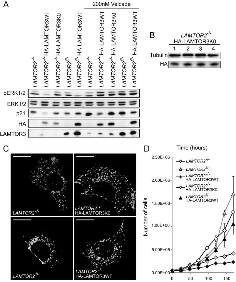
FIGURE 10.
Expression of HALAMTOR3K0 partially rescues LAMTOR2−/−MEF cell growth defect, but it does not restore late endosomal positioning. A, LAMTOR2−/−HA-LAMTOR3WT, LAMTOR2f/−HA-LAMTOR3WT, and LAMTOR2−/−HA-LAMTOR3K0 stable clones were generated as described under “Experimental Procedures.” LAMTOR2−/−, LAMTOR2f/−, LAMTOR2−/−HA-LAMTOR3WT, LAMTOR2f/−HA-LAMTOR3WT, and LAMTOR2−/−HA-LAMTOR3K0 MEF were treated with 200 nm Velcade for 7 h. Total cell lysates were separated by SDS-PAGE, analyzed by Western blotting, and probed with the indicated antibodies. B, LAMTOR2−/−HA-LAMTOR3K0 cells were harvested in 4 consecutive days. The lysates were separated by SDS-PAGE, analyzed by Western blotting, and probed with the indicated antibodies. C, LAMTOR2−/−, LAMTOR2f/−, LAMTOR2−/−HA-LAMTOR3WT, and LAMTOR2−/−HA-LAMTOR3K0 MEF were fixed in paraformaldehyde and subjected to indirect immunofluorescence analysis as described under “Experimental Procedures” using LAMP1 antibody. Epifluorescence pictures are shown. Bars, 20 μm. D, growth curves of LAMTOR2−/−, LAMTOR2f/−, and stable clones from LAMTOR2−/−HA-LAMTOR3WT, LAMTOR2−/−HA-LAMTOR3K0, and LAMTOR2f/−HA-LAMTOR3WT MEF. 5 × 10⁴ cells were plated into 10-cm dishes. The cells were counted using a Casy cell counter from Schärfe system GmbH at the indicated times (hours). The graph represents averages of three independent experiments (means ± S.D., n ≥ 3).

