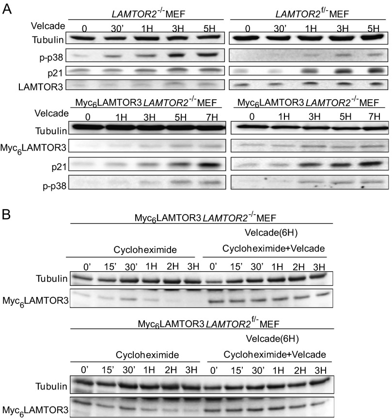
FIGURE 5.
The degradation of endogenous LAMTOR3 and Myc6LAMTOR3 is mediated by the proteasome. A, 80% confluent LAMTOR2−/−MEF, LAMTOR2f/−MEF, Myc6LAMTOR3LAMTOR2−/−, and Myc6LAMTOR3LAMTOR2f/− stable clones were treated with 50 nm Velcade for the corresponding time points. The cell lysates were separated by SDS-PAGE and probed with the indicated antibodies. B, Velcade and cycloheximide treatment of Myc6LAMTOR3LAMTOR2−/− and Myc6LAMTOR3LAMTOR2f/− stable clones. 80% confluent cells were pretreated with 50 nm Velcade for 6 h or left untreated. 50 μg/ml cycloheximide or 50 μg/ml cycloheximide plus 50 nm Velcade were then added to the cells for the corresponding time points. The cell lysates were separated by SDS-PAGE, analyzed by Western blotting, and probed with the indicated antibodies.

