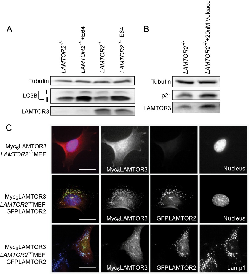
FIGURE 6.
The degradation of endogenous LAMTOR3 is lysosome-independent. A, LAMTOR2−/− and LAMTOR2f/−MEF were treated 100 μg/ml E64 for 7 h. Lysates were separated by SDS-PAGE, analyzed by Western blotting, and probed with the indicated antibodies. B, LAMTOR2−/− and LAMTOR2f/−MEF were treated with 20 nm Velcade for 7 h. The lysates were separated by SDS-PAGE, analyzed by Western blotting and probed with the indicated antibodies. C, Myc6LAMTOR3LAMTOR2−/− and Myc6LAMTOR3LAMTOR2−/−GFPLAMTOR2 MEF were subjected to indirect immunofluorescence analysis using antibodies against Myc and LAMP1. Epifluorescence pictures are shown. Because of the reduced levels of the protein and the diffuse cytosolic localization, the Myc6 panel shown for the Myc6LAMTOR3LAMTOR2−/− cells was acquired with a higher exposure time than the remaining images. Bars, 20 μm.

