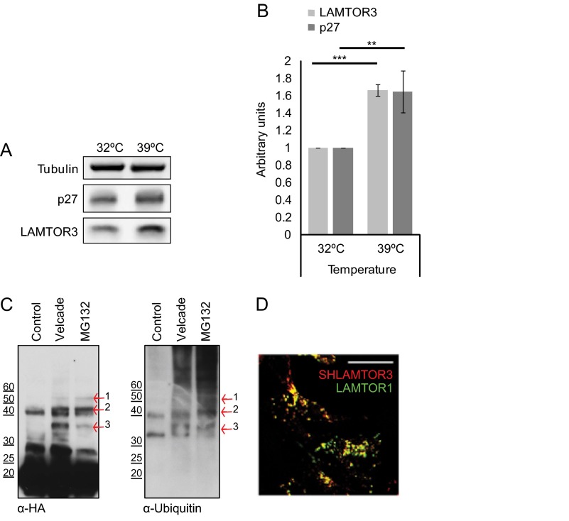
FIGURE 7.
The ubiquitin-mediated proteasomal degradation pathway controls LAMTOR3 degradation. A, the temperature-sensitive Ts20 cells derived from BALB/3T3 cells were kept at the permissive temperature of 32 °C or incubated for 16 h at 39 °C. Lysates were separated by SDS-PAGE, analyzed by Western blotting, and probed with the indicated antibodies. B, graphic shows the quantification of three independent experiments (means ± S.D., n = 3). **, p < 0.01; ***, p < 0.001. C, HEK293 SH-LAMTOR3 cells were induced with 400 ng/ml tetracycline for 24 h and supplemented with 200 nm Velcade or 10 μm MG132 for the final 7 h. The cells were lysed, and the obtained lysates were immobilized on a Strep-Tactin column. Samples were eluted with biotin, separated by SDS-PAGE, analyzed by Western blotting, and probed with the indicated antibodies. The arrows highlight bands positive for both HA and ubiquitin. D, HEK293 SH-LAMTOR3 cells were induced with 1 μg/ml tetracycline for 24 h and subjected to indirect immunofluorescence analysis using antibodies against HA and LAMP1. Confocal images are shown. Bar, 10 μm.

