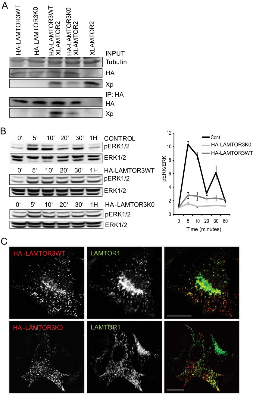
FIGURE 9.
Characterization of HA-LAMTOR3wt and HA-LAMTOR3K0. A, co-immunoprecipitation of XLAMTOR2 and HA-LAMTOR3wt or HA-LAMTOR3K0. HeLa cells were transfected with the corresponding constructs. 48 h after transfection, the cells were harvested, lysed, and subjected to immunoprecipitation (IP). The samples were loaded on SDS-PAGE, analyzed by Western blotting, and probed with the indicated antibodies. B, HeLa cells were mock transfected or transfected with HA-LAMTOR3wt or HA-LAMTOR3K0. The cells were serum-starved overnight and induced, 48 h after transfection, with 100 ng/ml EGF for the corresponding time points. Samples were then separated by SDS-PAGE and probed with the indicated antibodies. The graph represents averages of three independent experiments (S.D., n = 3). Cont, control. C, HeLa cells were transfected with HA-LAMTOR3wt or HA-LAMTOR3K0. The following day, the cells were split into coverslips and left to grow another 24 h. Transfected cells were fixed in paraformaldehyde, and immunofluorescence analysis was performed as described under “Experimental Procedures” using antibodies against HA and LAMTOR1. Bars, 20 μm.

