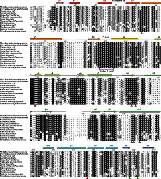
FIGURE 1.
Sequence alignment of type I PanKs from various pathogenic bacteria. MtPanK numbering and secondary structure are shown as defined by Das et al. (13). The blue triangle shows residue Arg-238, interacting with the phosphate bound in the P-loop, or inhibitory compounds. Red triangles show residues involved in pantothenate/phosphopantothenate binding. Green triangles show residues involved in adenine binding in the nucleoside binding site reported for Ec- and CbPanK. Sequences used in this alignment were as follows: MtPanK (UniProt ID P63810), Corynebacterium diphtheriae PanK (UniProt ID Q6NI48), EcPanK (UniProt ID P0A6I3), Salmonella typhi PanK (UniProt ID Q8Z318), Shigella dysenteriae PanK (UniProt ID Q32aF0), Klebsiella pneumoniae PanK (UniProt ID G0GSU7), Yersinia pestis PanK (UniProt ID Q8ZAN6), Haemophilus influenzae PanK (UniProt ID P44793), Vibrio cholerae PanK (UniProt ID Q9KV38), Coxiella burnetii PanK (UniProt ID Q83EV9), Bacillus subtilis PanK (UniProt ID P54556), Listeria monocytogenes PanK (UniProt ID Q8Y8I0), and Streptococcus pneumoniae PanK (UniProt ID Q97RH6).

