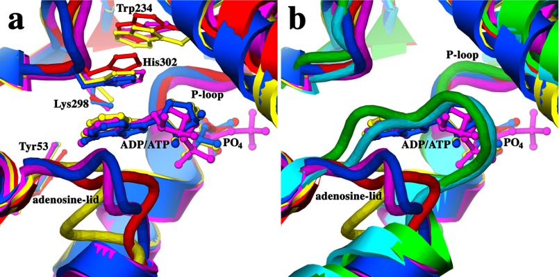
FIGURE 6.
a, superposition of MtPanK-2b-PO4 (red), EcPanK-AMPPNP (magenta, PDB code 1ESN), EcPanK-Pan-ADP (blue, PDB code 1SQ5), and CbPanK-ADP (yellow, PDB code 3TQC) showing the open form of the adenosine lid and conserved residues involved in adenosine binding. b, superposition of MtPanK-1a (green), EcPanK-CoA (light blue, PDB code 1ESM), MtPanK-2b-PO4 (red), EcPanK-AMPPNP (magenta, PDB code 1ESN), EcPanK-Pan-ADP (blue, PDB code 1SQ5), and CbPanK-ADP (yellow, PDB code 3TQC) showing the open and closed forms of the adenosine lid.

