FIGURE 2.
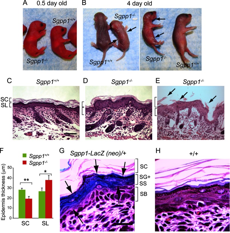
Sgpp1−/− pups exhibit focal desquamation and abnormal epidermal histology. A and B, Sgpp1+/+ and Sgpp1−/− mice at 0.5 days (A) and 4 days after birth (B). The arrows in B indicate sites of desquamation. C–E, histology of Sgpp1-deficient mouse skin. Paraffin sections of back skin from 2-day-old Sgpp1+/+ (C) and Sgpp1−/− (D and E) mice were stained with H&E. E represents a region of back skin undergoing peeling of the stratum corneum (marked by arrows). Bar, 200 μm. SC, stratum corneum; SL, subcorneal layer. F, stratum corneum and subcorneal layer thicknesses in 2-day-old Sgpp1+/+ and Sgpp1−/− mice. Bars represent mean values ± S.D. n = 4 for each genotype. Student's t test; *, p < 0.05; **, p < 0.01. G and H, activity of β-galactosidase, a surrogate marker for Sgpp1 expression, in 2-day-old mouse skin section. Sgpp1+/− (Sgpp1-LacZ (neo)/+) (G) and Sgpp1+/+ (+/+) (H) skin sections are shown. Arrows in G mark the keratinocyte layers that express β-galactosidase as a marker of Sgpp1 expression. This staining is absent in the +/+ skin section (H). Bar, 50 μm. SC, stratum corneum; SG, stratum granulosum; SS, stratum spinosum; SB, stratum basale.

