
FIGURE 4.
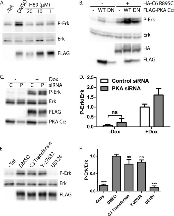
PKA inhibitor H89, but not dominant negative PKA Cα, PKA knockdown, or RhoA pathway inhibitors, blocks ERK activation by R895C TRPC6. A, M1R cells expressing R895C TRPC6 in a tetracycline-inducible manner were treated without (−Tet) or with tetracycline, followed by incubation with H89 at increasing concentrations or control carrier (DMSO). Phospho-ERK (P-Erk), ERK, and FLAG-TRPC6 expression were examined by Western blot of whole cell lysates. B, Western blot for phosphorylated ERK, ERK, HA, and FLAG of 293T cells transfected with HA-TRPC6 R895C and FLAG-PKA Cα wild type (WT) or dominant negative (DN) constructs, as indicated. C, R895C TRPC6-expressing cells were transfected with control (C) siRNA or siRNA targeting PKA Cα (P), followed by treatment with or without doxycycline to induce TRPC6 expression. Levels of phosphorylated and total ERK, FLAG-TRPC6, and endogenous PKA Cα were analyzed by Western blot. D, average normalized phospho-ERK/ERK levels in M1R cells expressing R895C TRPC6 under doxycycline treatment and transfected with the indicated siRNA. One-way ANOVA with Tukey's multiple comparison test was used; all pairwise comparisons with p < 0.001 except where indicated by ns (not significant); n = 3–7. E, tetracycline-inducible FLAG-TRPC6 R895C-expressing cells were treated without (−Tet) or with tetracycline, followed by incubation with carrier only (DMSO), Rho inhibitor (C3 transferase), ROCK inhibitor (Y-27632), or MEK inhibitor (U0126). Phospho-ERK and total ERK levels were analyzed by Western blot. F, average normalized phospho-ERK/ERK levels in cells treated as in E. One-way ANOVA with Dunnett's multiple comparison test was used versus DMSO; ***, p < 0.0001; ns, p > 0.05; n = 3–5. Error bars, S.E.