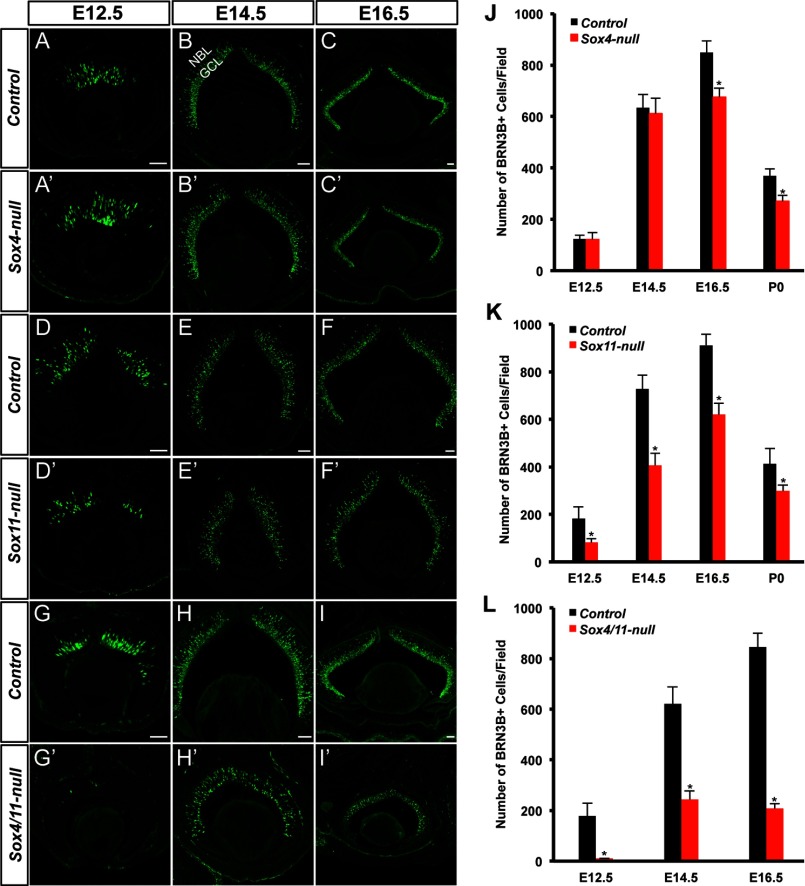
FIGURE 4.
Targeted disruption of Sox4 and Sox11 impairs the development of RGCs. A–I, A′–I′, cryo-sections of retinas from the control and mutant mice (Sox4-null, Sox11-null, and Sox4/11-null) at E12.5, E14.5, and E16.5 were immunolabeled with anti-BRN3B (green). J–L, quantification of RGCs in control and mutant mice at different embryonic stages. All experiments were repeated at least three times and error bars represent S.D. *, p < 0.01. GCL: ganglion cell layer, NBL: neuroblast layer. Scale bar: 50 μm.

