FIGURE 5.
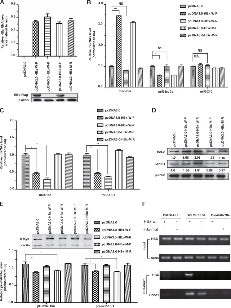
Induction of miR-15a/miR-16-1 down-regulation by HBx RNA. A, all four constructs expressed an equivalent amount of RNA (upper panel), but only the two protein-expressing constructs generated the expected FLAG-tagged HBx protein detected by Western blotting (lower panel). B, representative microRNAs responded to the expression of wild-type HBx protein (HBx-w-P) or RNA (HBx-wt-R) but not to the mutant HBx protein or RNA (HBx-m-P or HBx-m-R) in the miR-15a targeting site. C, the effects of HBx protein and RNA on the expression of miR-15a and miR-16-1 were quantified by real-time RT-PCR. D, the miR-15a/miR-16-1 cellular targets Bcl-2 and Ccnd-1 were only induced by wild-type HBx protein and RNA. The enhancing effects (quantified by scanning the protein gel) were quantified on the basis of the average of two independent experiments and are shown at the bottom of each gel. E, the effects of HBx protein and RNA on the expression of pri-miR-15a and pri-miR-16-1 were determined by real-time RT-PCR, and the degrees of c-Myc induction were quantified by Western blotting (top panel, top gel). F, capture of exogenous HBx and endogenous Ccnd1 transcripts with transfected biotinylated miR-15a but not bio-siGFP or bio-miR-20a. *, p < 0.05; **, p < 0.01; ***, p < 0.001; NS > 0.05 on the basis of experiments performed in triplicate.

