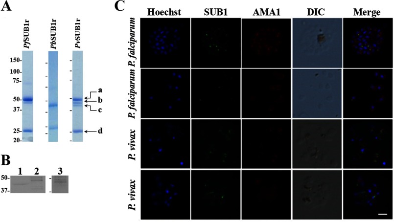
FIGURE 1.
Expression of PbSUB1r, PfSUB1r, and PvSUB1r and characterization of endogenous PvSUB1. A, Coomassie Blue staining of SDS-polyacrylamide gels of PbSUB1r, PfSUB1r, and PvSUB1r. Proteins were produced in baculovirus-infected insect cells, HPLC-purified, and concentrated (see “Experimental Procedures”). B, immunoblot of uninfected human erythrocytes (lane 1), P. falciparum 3D7 segmented schizonts (lane 2), and in vitro maturated P. vivax segmented schizonts (lane 3) probed with an anti-PvSUB1r serum. Molecular mass markers are in kDa. C, indirect immunofluorescence assays on air-dried P. falciparum 3D7 segmented schizonts (1st row of panels), individual merozoites (2nd row of panels), and P. vivax segmented schizonts (3rd and 4th rows of panels), probed with the anti-PvSUB1 polyclonal serum (green) and the anti-AMA1 rat monoclonal antibody 28G2 (red). Merozoite nuclei are labeled with Hoescht 33342. The scale bar represents 3 μm.

