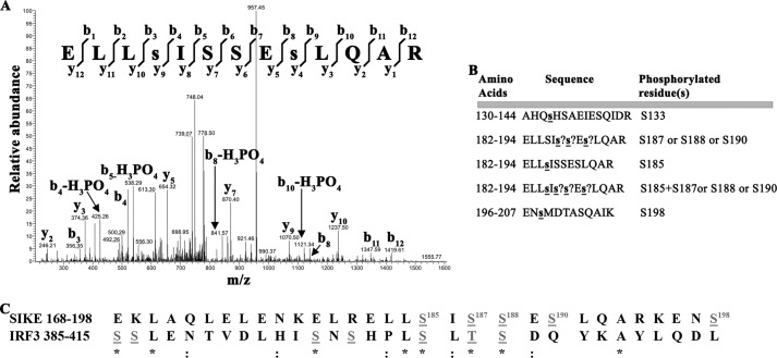
FIGURE 3.
TBK1 phosphorylates SIKE on six serine residues that mimic the IRF3 phosphorylation sequence. A, mass spectrum resulting from analysis of indicated phosphopeptide sequence. Two separate ion series were recorded simultaneously, the b- and y-ion series, which represent sequencing inward from the N and C termini, respectively. Lowercase s indicates phosphorylated residue. B, summary of phosphopeptide sequences identified using a Sequest search algorithm against custom databases generated from the His6-SIKE72 sequence. ? indicates that a phosphorylation site exists in peptide but cannot be uniquely assigned to a single serine residue. C, sequence alignment of known TBK1-mediated phosphorylation sites on IRF3, required for dimerization and activation, with TBK1-mediated phosphorylation sites on SIKE found through phosphopeptide mapping. Gray letters, phosphorylated residues; *, conserved site; :, functionally conserved site.

