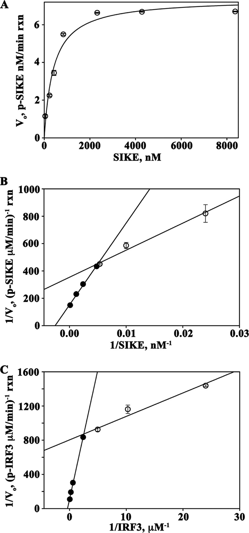
FIGURE 6.

SIKE is a substrate of TBK1. A, Michaelis-Menten plot of TBK1-mediated phosphorylation of SIKE with saturating ATP (100 mm), pre-activated TBK1 (4.93 nm), and SIKE varied from 0.043 to 8.4 mm. Data were fit to a 2-parameter rectangular hyperbola (SigmaPlot). B and C, Lineweaver-Burk plots of TBK1-SIKE assays with SIKE varied from 0.043 to 8.4 mm, ATP (100 mm) (B) and TBK1-IRF3 assays with IRF3 varied from 0.042 to 20.8 mm, ATP (100 mm) (C). TBK1 was at 4.93 nm in both assays. Data for 0.042–0.42 μm (○) and 1.7–20.8 μm (●) IRF3 and 0.043–0.23 μm (○) and 0.43–8.4 μm (●) SIKE were fit to a linear polynomial equation (SigmaPlot).
