
FIG. 1.
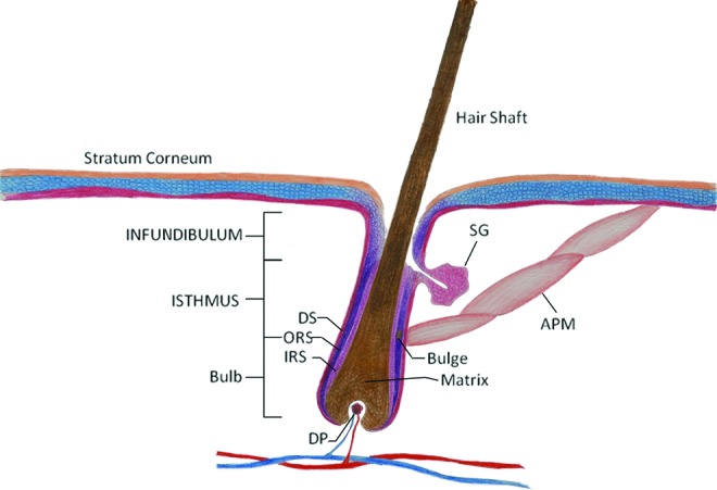
Schematic drawing of hair follicle. DS, dermal sheath; ORS, outer root sheath; IRS, inner root sheath; DP, dermal papilla; SG, sebaceous gland; APM, arrector pilli muscle. The illustration is not drawn in scale. Color images available online at www.liebertpub.com/teb