
Figure 1. Normal cardiac function in Epac1, Epac2 and DKO assessed by echocardiography.
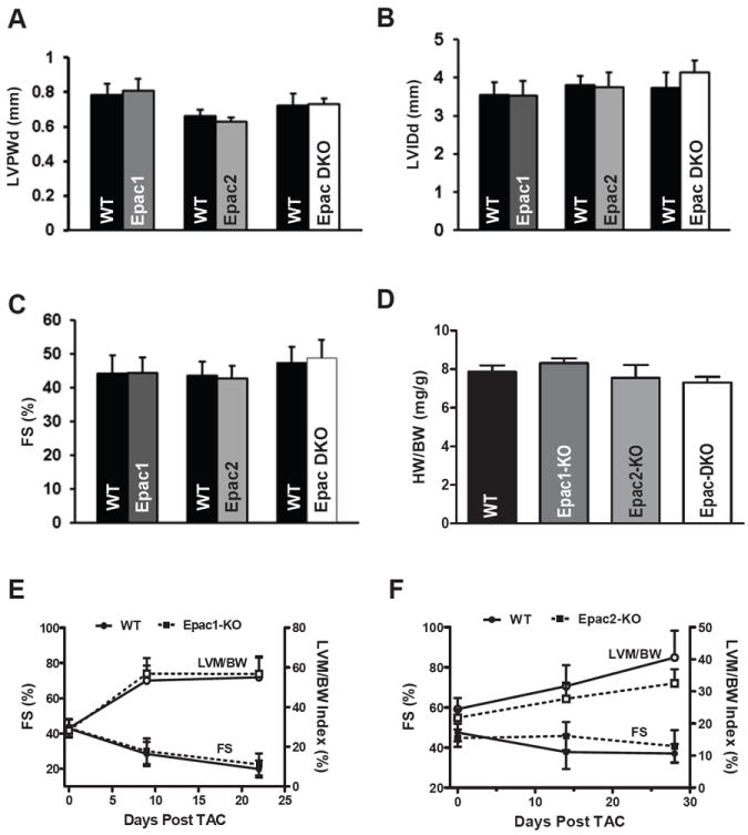
A. 8-months WT (n=6) and Epac1-KO (n=7) mice, 18-months WT (n=7) and Epac2-KO (n=6) mice and 18-months control (n = 7) and Epac-DKO (n=7) mice were measured by echocardiography. Between null mice and controls there was no difference in left ventricular wall thickness (LVPWd), left ventricle internal dimension size (LVIDd) at end of diastole (B), cardiac systolic function (C) or heart weight/Body weight ratio (D; Epac1-KO (n=3), Epac2-KO (n=5) and Epac-DKO (n=3)). E. Cardiac function (fractional shortening, FS) and hypertrophy (left ventricular mass/body weight; LVM/BW) in Epac1-KO (n= 8) and (F) Epac2-KO (n=9) vs. WT measured during 3-4 weeks after TAC.