
Figure 4. Epac2-KO deletion reduced ISO-dependent increase of pacing-induced ventricular tachycardia.
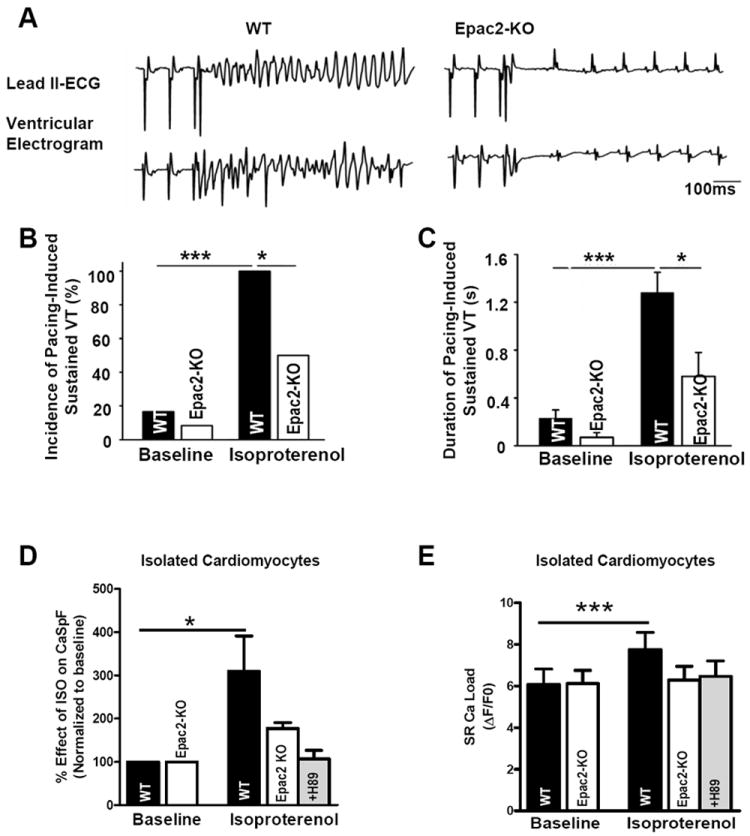
A. Representative simultaneous surface ECG (lead 2) and intra-cardiac ventricular electrogram after ISO (0.5mg/kg) revealed sustained ventricular tachycardia (SVT) in WT and sinus rhythm in Epac2-KO after S1-S2 extra-stimuli at pacing CL of 90ms. B. Mean SVT incidence before and after ISO stimulation. C. Mean duration of SVT under same conditions (n=12 WT and 12 Epac2-KO mice for C and D). D. Percent effect of ISO on CaSpF in isolated cardiomyocytes from WT (n=7) and Epac2-KO±H89 (n=5 and n=12). E. SR Ca load before and after ISO in same cells than D. *P<0.05, ***P<0.01.