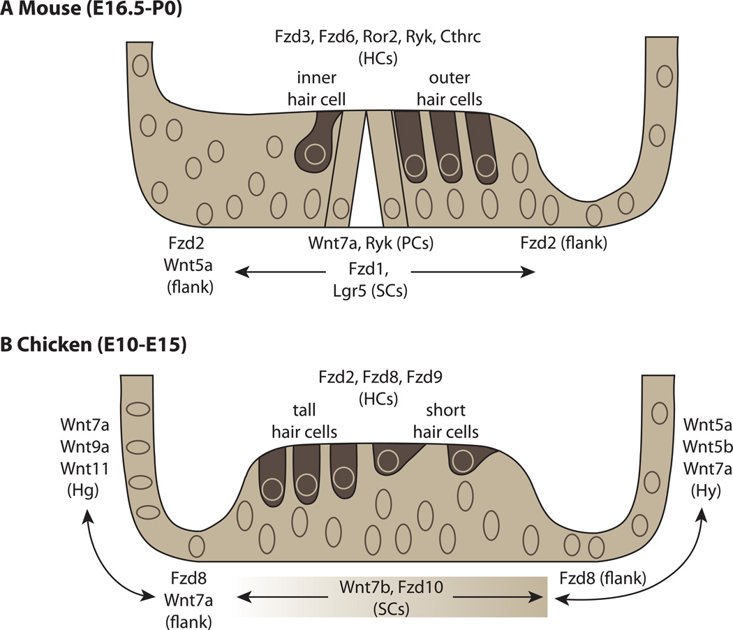
Figure 3.
Expression patterns of Wnt ligands and receptors in the mouse and chick cochlear ducts.(A) Expression in the mouse cochlea from E16.5-P0. Fzd3, Fzd 6, Ror2, Ryk and Cthrc are expressed in hair cells (HCs) [64, 66, 83] while Fzd1 and Lgr5 are expressed in support cells (SCs) [65, 35, 36]. Wnt5a is expressed in the greater epithelial ridge on the neural side of the organ of Corti, while Fzd2 flanks the hair cells on both sides of the organ [65, 67]. (B) Expression in the chick cochlea from E10-E15 [39]. Fzd2 and Fzd9 are high in hair cells while Fzd8 is much weaker in comparison to its expression on the immediate flanks of the sensory organ in clear cells (on the neural side) and border cells (on the abneural side). Wnt7b and Fzd10 are expressed in the SCs in a gradient with stronger expression on the abneural side. Several Wnts are expressed in non-sensory cells types including the homogene cells (Hg) and the hyaline cells (Hy). To focus on regional or cell-type-specific expression patterns, Wnt receptors/ligands/inhibitors with ubiquitous and/or exceptionally weak expression at these time points have been omitted. Details of omitted genes can be found in [39].

