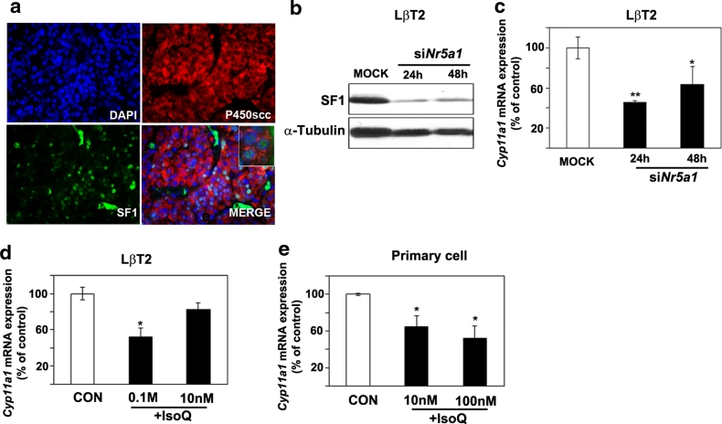
Fig. 7.
Co-localization of P450scc and SF-1 in rat PAs and reduction of Cyp11a1 expression following down-regulation or inhibition of SF-1 in gonadotroph cells. a Immunofluorescent staining with antibodies against P450scc (red) and SF-1 (green) was performed on FFPE pituitary tumor tissues from MENX mutant rats. Nuclei were counterstained with DAPI. The inset in MERGE shows the co-localization of SF-1 and P450scc in rat tumor cells. Original magnification: ×200; inset: ×400. b LβT2 cells were transfected with scrambled (MOCK) or siRNA oligos against the mouse Nr5a1 gene (siNr5a1). SF-1 and α-tubulin expression levels were assessed by Western blotting 24 and 48 h after transfection. c In samples parallel to “b”, Cyp11a1 expression level was assessed by qRT-PCR as indicated in the legend of Fig. 1, and is reported relative to the expression level in mock-transfected cells arbitrarily set to 100. d LβT2 cells were treated with different concentrations of the SF-1 inhibitor IsoQ (0.1 M and 10 nM) or left untreated (CON). After 24 h, we determined Cyp11a1 expression levels as in “c”. e Primary pituitary tumor cells from mutant rats (n = 3) were incubated with IsoQ as in “d”. After 24 h, we assessed Cyp11a1 expression levels by qRT-PCR as in “c”. Data from primary cultures were analyzed independently with six replicates each and were expressed as the mean ± SEM. *P < 0.05, ***P < 0.001 versus MOCK or CON

