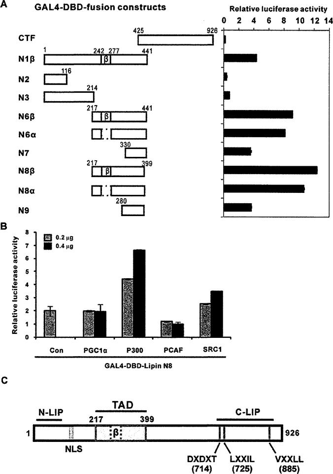
Figure 4. GAL4-DBD fusion proteins containing the lipin1 TAD activate UAS-driven transcription.
(A) Left-hand panel, pFA-CMV-lipin1 constructs encoding GAL4-DBD fusion proteins with various fragments of lipin1 and the reporter vector pFR-luciferase were introduced into HEK-293 cells. Right-hand panel, luciferase activity was measured 48 h after transfection. (B) NIH 3T3 cells were co-transfected with pFR-Luc and GAL4-DBD-lipin1 TAD fusion constructs (GAL4-DBD–lipin1 N8) along with co-activator plasmids (PGC-1α, P300, PCAF and SRC-1). Luciferase activity was normalized to Renilla luciferase and the data shown are representative of three independent experiments performed in triplicate. (C) Schematic diagram depicting the domains and motifs of the human lipin1. N-LIP and C-LIP indicate evolutionarily conserved N-terminal and C-terminal lipin domains. TAD indicates the transcriptional activation domain delineated in the present study. The nuclear localization signal (NLS), PAP enzymatic motif (DXDXT) and nuclear receptor interaction motifs (LXXIL and VXXLL) are also shown.

