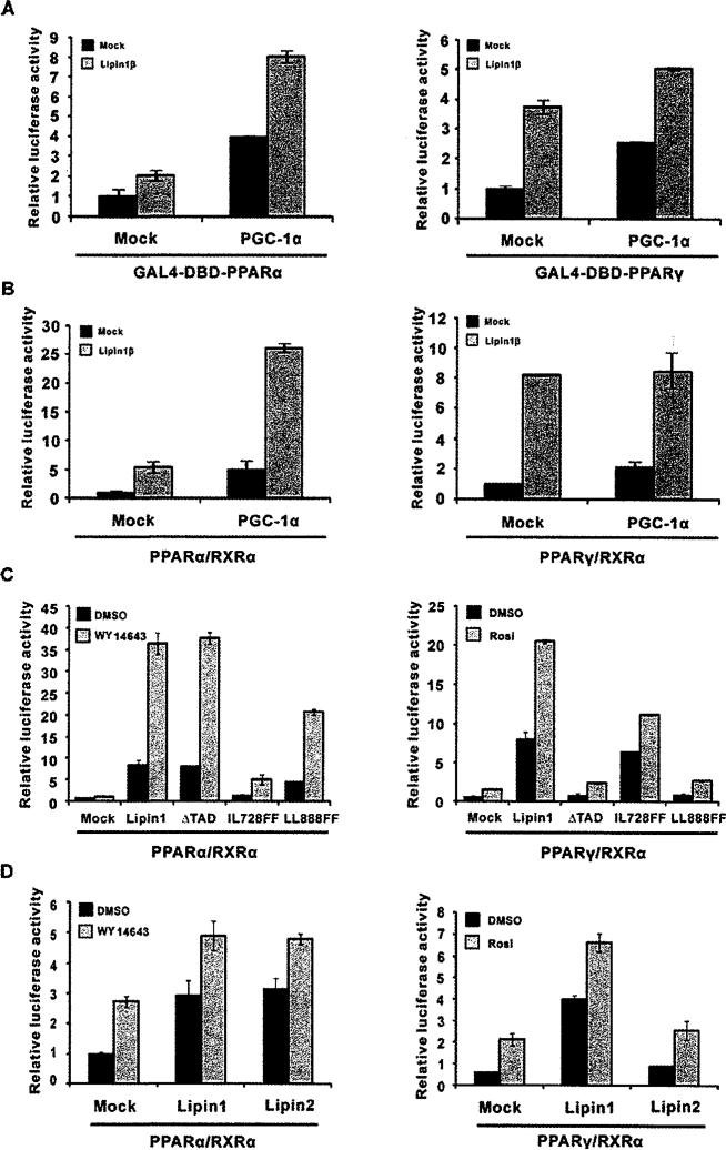
Figure 5. The mechanism of lipin1-mediated PPARγ activation differs from PPARα activation.
(A) Expression vectors for GAL4-DBD–PPARα (left-hand panel) or the GAL4-DBD–PPARγ (right-hand panel) fusion proteins and pFR-Luc were transfected into NIH 3T3 cells along with pcDNA-PGC-1α and/or pSG5-lipin1, as indicated. (B–D) PPRE-tk-luciferase and expression vectors for PPARα (left-hand panels), PPARγ (right-hand panels), RXRα, PGC1α, lipin1, lipin1 mutants (ΔTAD, LL728FF and LL888FF) or lipin2 were transfected into NIH 3T3 cells as indicated. Rosi., rosiglitazone.

