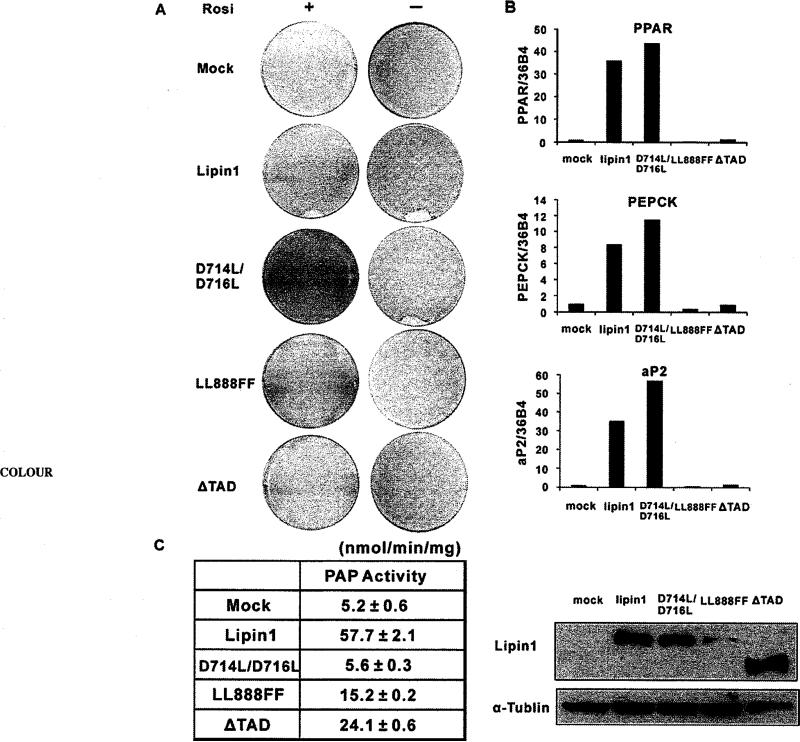
Figure 6. Lipin1 mediates differentiation of 3T3-L1 pre-adipocytes through its PPARγ-co-activating activity.
3T3-L1 cells were transfected with pSG5-KF2M1 (Mock), pSG5-lipin1 and lipin1 mutants (D714L/D716L, LL888FF and ΔTAD) using electroporation and induced to differentiate when cells reached below 70% confluncy. (A) Oil Red O staining of transfected cells on day 7 after induction of differentiation with (+) or without (–) 1 μM rosiglitazone. Forced expression of lipin1 and mutants were detected at day 2 after induction of differentiation. (B) The expressions of adipocyte-specific genes were analysed by real-time PCR at day 7 after differentiation in the presence of 1 μM rosiglitazone. Relative mRNA levels of each gene were normalized to the levels of 36B4 mRNA. The data shown are representative of three independent experiments performed in triplicate. (C) PAP1 activity was measured with the extracts from HEK-293 cells which overexpress wild-type or mutant lipin1 (left). The data shown are representative of three independent experiments performed in triplicate. Western Blots of wild-type or mutant lipin1 are shown (right).

