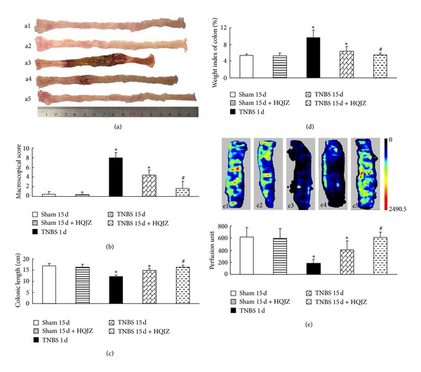
Figure 1.

Macroscopic observation and colonic blood flow. (a) Representative images of colon, a1: Sham 15 d; a2: Sham 15 d + HQJZ; a3: TNBS 1 d; a4: TNBS 15 d; a5: and TNBS 15 d + HQJZ. (b) Macroscopic scores. (c) Colonic length. (d) Weight index of colon. (e) Representative images and quantitative analysis of colonic blood flow, e1: Sham 15 d; e2: Sham 15 d + HQJZ; e3: TNBS 1 d; e4: TNBS 15 d; and e5: TNBS 15 d + HQJZ. The magnitude of colonic blood flow is represented by different colors, with blue to red denoting low to high. Data were mean ± SEM (n = 8). *P < 0.05 versus Sham group; # P < 0.05 versus TNBS 15 d group.
