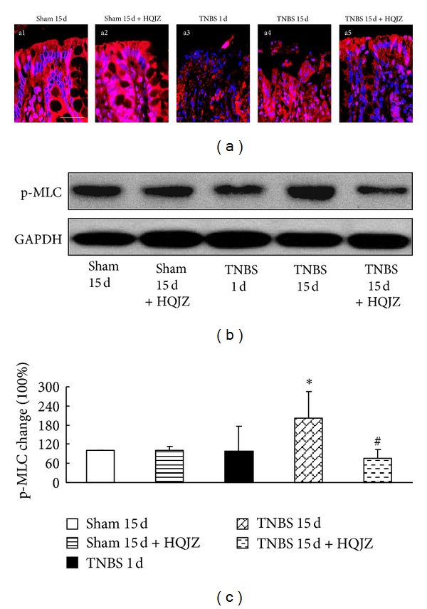
Figure 7.

Distribution of F-actin in colonic mucosa and Western blot analysis of p-MLC. (a) Distribution of F-actin in colonic mucosa. The red zone represents the distribution of F-actin, and the blue zone represents nuclei. Bar = 25 μm. (b) Representative Western blots of p-MLC and GAPDH. (c) Quantitative analysis of p-MLC proteins. Data were mean ± SEM (n = 5). *P < 0.05 versus Sham group; # P < 0.05 versus TNBS 15 d group.
