Fig. 3.
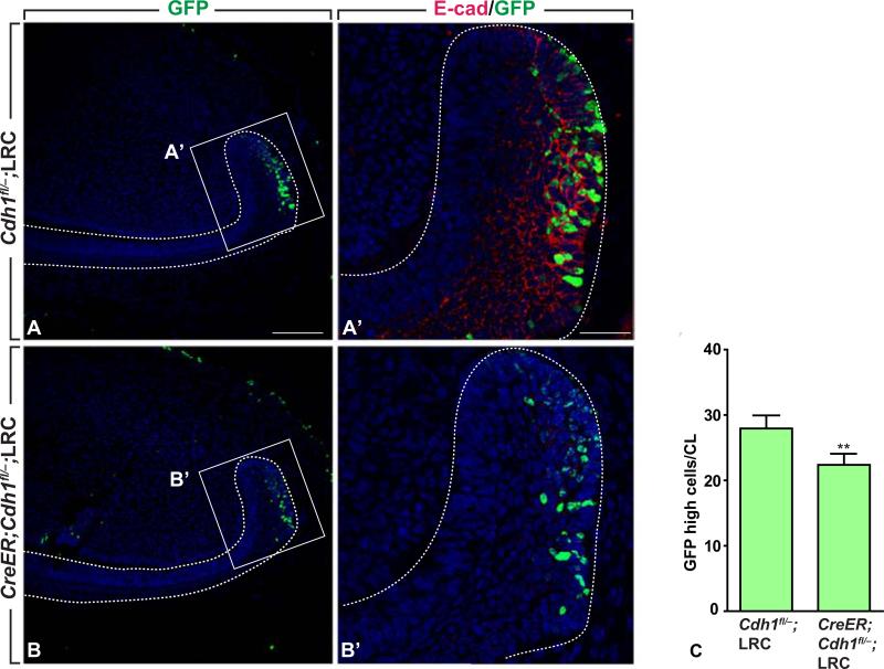
Decreased numbers of GFP-high LRCs after deletion of E-cadherin in the cervical loops. (A, A’) Incisor section of control and E-cadherin mutant stained with GFP antibody. (B, B’) Incisor section of control and E-cadherin mutant stained with GFP and E-cadherin antibodies. (C) Quantification of GFP-high LRCs in controls and E-cadherin mutants; mean ± SEM (n=3; p<0.05). Dotted lines outline the labial cervical loops. Scale bars: 100 μm in A, C; 50 μm in B, D.

