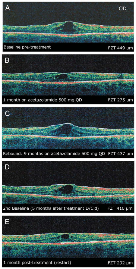
Fig. 1.
Patient 1. A, Baseline OCT image (90°) of the right eye showing CME. B, Optical coherence tomography image of the same eye after 1 month of treatment with acetazolamide. C, Optical coherence tomography image of the same eye 9 months later showing a rebound of macular cysts from baseline. Acetazolamide was discontinued at this time. D, Optical coherence tomography image of the same eye 5 months after discontinuing treatment. Treatment with acetazolamide was restarted at this time. E, Optical coherence tomography image of the same eye 1 month post retreatment demonstrating a reduction in size of the macular cysts. All images were obtained on a Stratus system.

