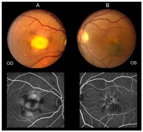
Fig. 1.
Fundus photograph obtained at the most recent follow-up visit of the right eye (A) demonstrating a vitelliform-appearing macular lesion and left eye (B) demonstrating macular hypopigmentary changes, RPE mottling, and a flat choroidal nevus inferior to the fovea. Fluorescein angiogram (late frames) of both eyes shows window defects and late staining in the macula of each eye with pooling of fluorescein dye in the macula of the right eye as well as hypofluorescent loci in each eye.

