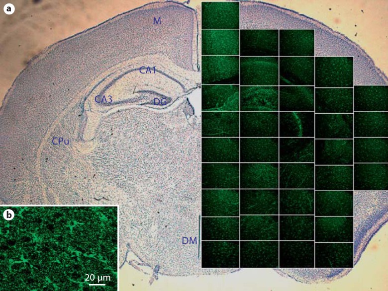
Fig. 2.
Distribution of activated kappa opioid receptors in a wild-type P9 posterior brain. Phospho-specific labeling of KOR-P in a posterior brain 30 min after stimulation with U50,488. a Spatial representation of a coronal slice of brain containing the hippocampus and hypothalamus. Photomicrograph magnification 400X. Brain regions are identified in the contralateral hemisphere: M = motor cortex; DG = dentate gyrus; CAI field = CAI of hippocampus; CA3 field = CA3 of hippocampus; CPu = caudate putamen (striatum); DM = dorsal medial nuclei of the hypothalamus. b Enlargement of an individual micrograph showing specificity of KOR-P labeling to cell bodies and processes in the motor cortex. Sections with the primary antibody deleted showed no KOR-P immunoreactivity

