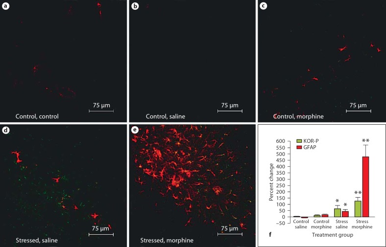
Fig. 4.
KOR-P and GFAP immunoreactivity increases in the molecular layer of the hippocampus in wild-type neonatal mice exposed to stress and treated with morphine. The distribution of GFAP immunoreactivity (red) and activated KOR (green) is compared in the molecular layer of the hippocampus from 5 treatment groups: untreated control (a), saline-injected control (b), morphine-injected control (c), stressed with saline injections (d), and stressed with morphine injections (e). Photomicrograph magnification 400 X. f Percent change in pixel intensity for both GFAP and KOR-P in the molecular layer of the wild-type mouse hippocampus. Red bars represent the percent change in GFAP immunoreactivity, and green bars represent the percent change in KOR-P immunoreactivity in the treatment groups compared to the untreated controls. * p < 0.05; < p < 0.01.

