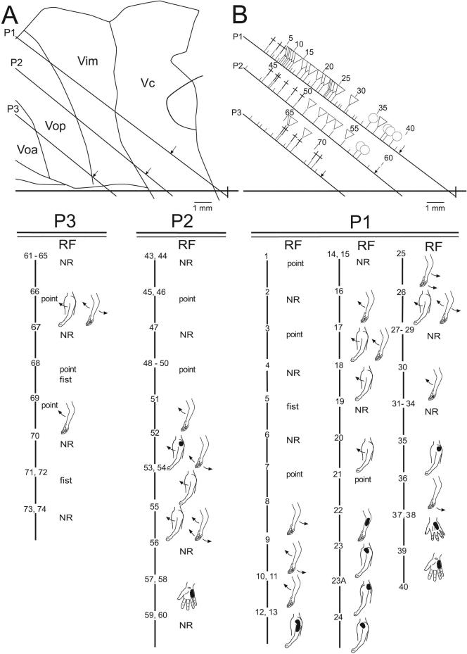
Figure 1.
Sagittal map of thalamic neuronal location relative to the AC-PC line (horizontal line, PC as indicated) and nuclear boundaries, in the patient with psychogenic dystonia. Electrode trajectories (P3, P2 and P1) are shown by the oblique lines. Locations of recording sites are indicated by ticks to the right of each trajectory. Long tics indicate location of a neuron which responded during sensory stimulation, or active movement, or both; short tics indicate unresponsive neurons. The symbol at a long tic indicates the presence of a neuron responding during active movement (cross), cutaneous stimulation (circle), and deep stimulation (triangle), as described in the methods. Sites are numbered sequentially and numbers are indicated for every fifth tick. Scale as indicated.
Lower panels P1, P2, and P3 show the site number, and receptive field (RF). RF is indicated to the right and below the site number. Note that many cells responded to movement of two joints. For example, one cell responded to extension of both shoulder and elbow. Shading on the figurine indicates the part of the body where mechanical stimulation-evoked cellular activity. All shaded figurines indicate cutaneous stimulation, except for neurons 22 - 24, 35, at which cellular activity was evoked by manipulation of muscle but not by manipulation of the overlying skin. The maximum size of the representation of a part of the body was estimated from the lengths of single electrode trajectories along which all RFs included a single joint, as in a previous study 7. For example, in electrode trajectory P2, passive shoulder movements were represented by neurons 52 to 55, a distance of 2.5 mm.

