Figure 3.
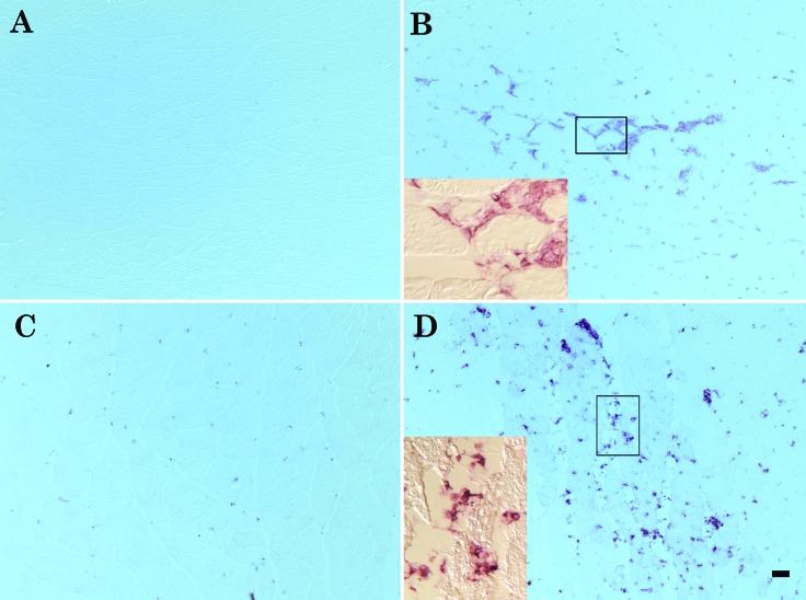
F4/80 and CD68 immunolabeling. (A) Uninjured lateral gastrocnemius muscle is negative for F4/80 staining. (B) Extensive invasion of lateral gastrocnemius muscle by F4/80-positive cells at 48 h after injury. (C) CD68-positive macrophages are present in the uninjured lateral gastrocnemius muscle. (D) CD68-positive macrophages increase in area percentage and size in the injured lateral gastrocnemius muscle at 48 h after injury. Bar, 50 µm. Insets at higher magnification (20×) show the positive cells surrounding and infiltrating the injured fibers.

