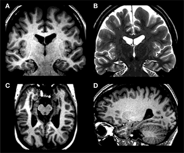
Figure 1.

Bilateral hippocampal atrophy in ML with T1-weighted images [(A) coronal; (C) axial; (D) sagittal] demonstrating reduced hippocampal size in all directions – in the absence of marked extrahippocampal atrophy; T2 weighted coronal image (B) demonstrating bilateral loss of internal structure – here: a further marker of bilateral hippocampal atrophy. On clinical MRIs left side of the image is right side of the patient.
